


Mesh Complications Patient Story 13
Leg & Groin Pain after TOT sling placement
Mesh Complications: Stress incontinence, persistent left leg & groin pain.
Treatment: Removal of sling and simultaneously replace with TVT sling.
The patient is a 53- year- old female who has undergone the following surgeries for stress urinary leakage:
- 1991 - Anterior Repair
- 1999 - Pubovaginal Cadaveric Sling
- 2002 - 2nd Pubovaginal Cadervic Sling
- 2011 - TOT Obtryx
The patient has experienced groin pain and inner thigh pain since her TOT surgery for a period of four weeks. The patient reported the day after surgery her pain was a ten on a pain scale ranging from 0 (no pain) to ten (worst pain possible). The patient did report a decrease in her pain at times, however, stated, “It can still be debilitating.” She came to International Urogynecology Associates for evaluation and treatment.
Upon examination, the patient had reproducible pain just by faintly touching the sling. A cough test confirmed stress urinary incontinence. After an informed consent, she elected to proceed with surgery. During the area of the exit wound was readily identifiable and is marked. (Figure 1)
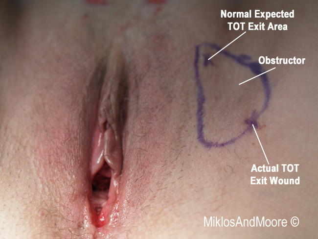
Figure 1
Figure 1: The patient is marked to show the obturator fossa (the oval circle). The upper X is the normal area of entry for a TOT sling. The lower X is the exit wound for this patient’s TOT sling. This low area of exit allows for the sling to cross the crease of the leg and in theory is responsible for the persistent leg pain.
The patient was taken to surgery and under IV sedation the sling was removed. The skin was mobilized until the sling was identified and the sling was dissected away from the urethra and bladder. The sling was divided in the midline and dislodge from the patients left side. A firm pulling allowed the slings release and removal.

Figure 2
Figure 2: TOT sling removed from the left side only.
The patient had a dramatic reduction in pain. Placing a TVT sling during surgery also treated the patient’s urine leakage. She is doing well post operatively and is without leakage.
Click here to find out more about TOT Sling complications.
Click here for related patient stories